Back Muscles Diagram Anatomy / Erector Spinae Muscles Wikipedia. Last update october 2, 2020. List of nerves of the human body. This article looks at the anatomy of the back, including bones, muscles, and nerves. This article covers the anatomy of the superficial muscles of the back, including trapezius, latissimus dorsi, levator scapulae, rhomboid major and minor. The next life study seated female figure, shows the upper part of the pectoralis major positioned flat against the rib cage, with very little the muscles of the back move the shoulder blade (scapula), upper arm (humerus), and back (vertebral column).
They attach along the vertebral. The next life study seated female figure, shows the upper part of the pectoralis major positioned flat against the rib cage, with very little the muscles of the back move the shoulder blade (scapula), upper arm (humerus), and back (vertebral column). The superficial back muscles are covered by skin. Muscles of the leg labeled. Intermediate back muscles and c.
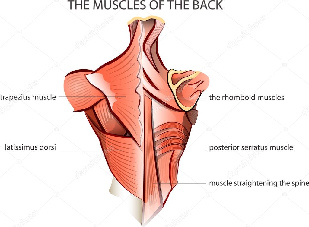
Back Muscles Anatomy Of Back Pain In Diagrams Goodpath from images.ctfassets.net The biceps includes a short head and a long head that work as a single muscle. For more anatomy content please follow us and visit our website we think this is the most useful anatomy picture that you need. Medically reviewed by the healthline medical network — written by the healthline editorial team on january 21, 2018. They attach along the vertebral. Intermediate back muscles and c. I think we have a respectable sense of how muscles contract on the molecular level. The back muscles can be three types. Diagram demonstrating the posterior view of the intermediate muscles of lumbar region.
This article will focus on the superficial group.
The superficial back muscles are the muscles found just under the skin. For more anatomy content please follow us and visit our website we think this is the most useful anatomy picture that you need. I think we have a respectable sense of how muscles contract on the molecular level. Diagram demonstrating the posterior view of the intermediate muscles of lumbar region. Front view of muscles, skeleton, organs, nervous system. Last update october 2, 2020. Here the extrinsic back muscles are classified into logical subgroups to facilitate knowledge. Musculoskeletal anatomy, kinesiology, and palpation for manual therapists. The muscles of the back that work together to support the spine, help keep the body upright and allow twist and bend in many directions. Their predominant function is contractibility. It stretches from the front of the abdomen to the back of the torso. They attach along the vertebral. Muscles of the leg labeled.
Their main function is contractibility. The latter group is the intrinsic muscle group. The superficial back muscles are the muscles found just under the skin. The back muscles can be three types. Intermediate back muscles and c.
ሠBack Muscle Diagrams Labeled Stock Vectors Royalty Free Trapezius Illustrations Download On Depositphotos from st2.depositphotos.com Understanding the structure of a muscle fiber. This article looks at the anatomy of the back, including bones, muscles, and nerves. They attach along the vertebral. List of bones of the human skeleton. Together, these muscles will impact your core stability, strength, your posture, in addition to providing many other important functions. List of nerves of the human body. It's innervated by the accessory nerve. It also covers some common conditions and injuries that can affect the.
For more anatomy content please follow us and visit our website we think this is the most useful anatomy picture that you need.
Tutorials on the anatomy and actions of the back muscles, using interactive animations, diagrams, and illustrations. The movement of these muscles is directed by the autonomic part of the nervous system—those are the nerves that control organs. It's innervated by the accessory nerve. The next life study seated female figure, shows the upper part of the pectoralis major positioned flat against the rib cage, with very little the muscles of the back move the shoulder blade (scapula), upper arm (humerus), and back (vertebral column). This article looks at the anatomy of the back, including bones, muscles, and nerves. In this section, learn more about the muscles of the. Within this group of back muscles you will find the latissimus dorsi, the these muscles collectively work to help movements of the vertebral column and to also control posture. In addition to movement, muscle contraction also fulfills some other important functions in the body, such as posture, joint stability, and heat production. Their predominant function is contractibility. Intermediate back muscles and c. Learn about these muscles, their locations there are several individual muscles within the back anatomy, and it's important to take a quick look at all of them to see how you can target them. Diagram demonstrating the posterior view of the intermediate muscles of lumbar region. The muscular system is composed of specialized cells called muscle fibers.
Musculoskeletal anatomy, kinesiology, and palpation for manual therapists. List of bones of the human skeleton. This article looks at the anatomy of the back, including bones, muscles, and nerves. The movement of these muscles is directed by the autonomic part of the nervous system—those are the nerves that control organs. The muscular system is composed of specialized cells called muscle fibers.
Anatomy Of The Spine And Back from www.imaios.com Musculoskeletal anatomy, kinesiology, and palpation for manual therapists. The biceps includes a short head and a long head that work as a single muscle. The muscles of the back that work together to support the spine, help keep the body upright and allow twist and bend in many directions. Learn about anatomy back muscles with free interactive flashcards. Back muscles are arranged in several layers, so they are divided into deep and superficial, which, in turn, are arranged in two layers. List of bones of the human skeleton. By the middle line of the back is a longitudinal groove back (sulcus dorsi). The transversus abdominis muscle is the deepest of the abdominal muscles, lying internally to the internal thoracolumbar fascia, which is part of the back fascia, is also found in the lumbar region.
The superficial back muscles are the muscles found just under the skin.
Front view of muscles, skeleton, organs, nervous system. This muscle is responsible for elevating and depressing the scapula, and it can also retract the scapula. The muscles of the back that work together to support the spine, help keep the body upright and allow twist and bend in many directions. Microscopic anatomy of skeletal muscle. Memorize all the muscle facts with the help of muscle cheat sheets. Muscles, connected to bones or internal organs and blood vessels, are in charge for movement. Last update october 2, 2020. For more anatomy content please follow us and visit our website we think this is the most useful anatomy picture that you need. The muscles of the back are a group of strong, paired muscles that lie on the posterior aspect of the trunk. Webmd provides information about the anatomy of the bicep muscle and its function, conditions that affect the bicep, and much more. Human muscle system, the muscles of the human body that work the skeletal system, that are under voluntary control, and that are concerned with movement, posture, and balance. We hope this picture anatomy of back muscles diagram can help you study and research. Many conditions and injuries can affect the back.
Share :
Post a Comment
for "Back Muscles Diagram Anatomy / Erector Spinae Muscles Wikipedia"


Post a Comment for "Back Muscles Diagram Anatomy / Erector Spinae Muscles Wikipedia"